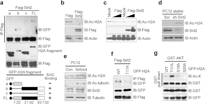
Figure 6. Akt recruits Sirt2 for H2A deacetylation.
(a) 293T cells were co-transfected with FLAG-SIRT2 and GFP-H2A fragments. Cell extracts were immunoprecipitated with anti-FLAG antibody and immunoblotted with the antibodies indicated. Schematic representation of H2A fragments and constructs used to identify the SIRT2 interaction region in H2A (lower). (b) 293T cells were transfected with the indicated plasmids for 24 h, and equal amounts of cell lysate were subjected to immunoblotting using the indicated antibodies. (c) PC12 cells were transfected with Mock and FLAG-SIRT2 constructs in a dose-dependent manner. The cell extracts were immunoblotted with anti-acetyl H2A (K5) antibodies as indicated. (d) PC12 cells with stable knockdown of SIRT2 were harvested and lysed. Proteins were immunoblotted with anti-acetyl H2A (K5). (e) PC12 cells were treated with Sirtinol (50 μm for 24 h) and immunoblotted with the indicated antibodies. (f) PC12 cells were co-transfected with FLAG-SIRT2 and GFP-H2-WT (or H2A-T17A) constructs. The cell extracts were immunoprecipitated with anti-FLAG antibody and immunoblotted with the antibodies indicated. (g) PC12 cells were co-transfected with GST-Akt and GFP-tagged H2A constructs (WT, T17A, S19A, and S20A). Proteins were pulled down with GST resin and visualized by immunoblotting.

