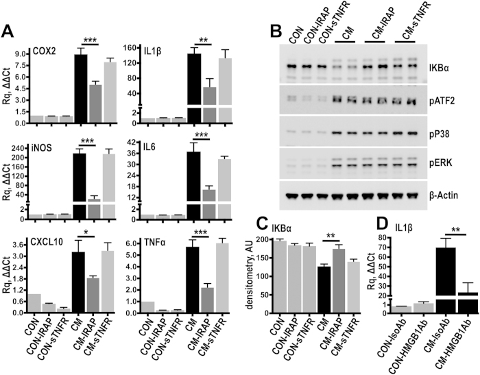
Figure 6. Interleukin 1 is the dominant inflammatory factor in the injury conditioned medium.
(A) Resting fibroblasts were stimulated for 3 hours with injury conditioned medium (CM) supplemented with 20 ng/ml of either IRAP or sTNFR. Control cells were kept in plain culture medium (CON) and additional controls were incubated with IRAP (CON-IRAP) or sTNFR (CON-sTNFR). Gene expression was measured by TaqMan qPCR. ACTB served as the housekeeping gene. N = 4 independent experiments. (B,C) To examine acute signalling activation cells were stimulated for 25 minutes with conditioned medium supplemented either with IRAP or sTNFR and signalling activation was examined by immunoblotting (B). IκΒα levels were quantified by densitometry (C) N = 3 independent experiments. (D) Resting cells were stimulated with injury conditioned medium which was previously treated for 2 hours with HMGB1 antibodies (HMGB1Ab; 5 μg/ml) to neutralize soluble extracellular HMGB1, or with isotype control antibodies (IsoAb; 5 μg/ml). IL1β expression was measured by TaqMan qPCR. N = 6 independent experiments. *P ≤ 0.05, **P ≤ 0.01, ***P ≤ 0.001; Anova with Fisher’s LSD multiple comparison test.

