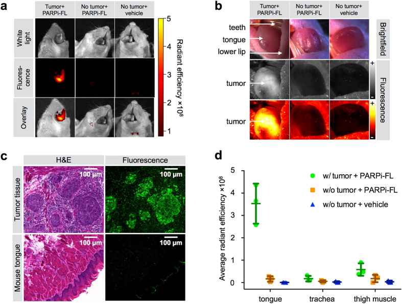
Figure 5. Detection of an orthotopic OSCC xenograft model in mouse tongue.
(a) Epifluorescence imaging of tumor-bearing tongues (FaDu) and healthy mouse tongues from animals injected with PARPi-FL (75 nmol/167 μl PBS with 30% PEG300) or vehicle (PBS with 30% PEG300) 90 minutes prior to imaging. (b) Imaging with a Lumar fluorescence stereoscope. Images were taken in brightfield and with 488 nm laser excitation. Fluorescent images are displayed in grayscale and intensity-scaled (“+” = maximum signal, “−” = minimum signal). (c) Cryopreserved tongue sections were imaged with a confocal microscope to detect PARPi-FL following fixation. H&E staining in adjacent sections for anatomical and pathological evaluation of PARPi-FL localization. (d) Average radiant efficiency [p/s/cm2/sr]/[μW/cm2] of PARPi-FL in excised tumor-bearing and healthy tongues as well as trachea and thigh muscle. Means and SD from n = 3 animals/group are displayed.

