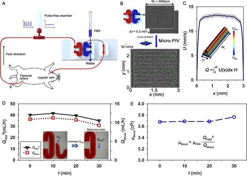
Figure 1. Experimental system for ex vivo monitoring of biophysical properties.
(A) Blood is supplied into the extracorporeal fluidic network by connecting an extracorporeal loop to the blood vessels of a rat model. The extracorporeal loop consists of a pulse-free chamber (air cavity = 1.5 mL), and H-shaped and straight microchannels. The syringe and microfluidic devices were illustrated by the authors using SolidWorks software (Dassault Systèmes SolidWorks Corp., USA). (B) Procedure of micro-PIV measurement. The H-shaped microchannel has a width (W1) of 3000 μm and a height (H) of 80 μm. An instantaneous velocity field superimposed on the corresponding flow image is represented in the inset. (C) Axial velocity profile along the lateral position (x) of the channel. 3D velocity distribution in the channel and calculation of flow rate (Q). (D) Temporal variations in the flow rate of the PBS solution at the hydrodynamic balancing state (QPBSB) and blood flow rate (QBlood) measured by using Eq. (2). Microscopic images show hydrodynamic unbalancing and balancing states in the H-shaped microchannel. (E) Temporal variation of blood viscosity (μBlood).

