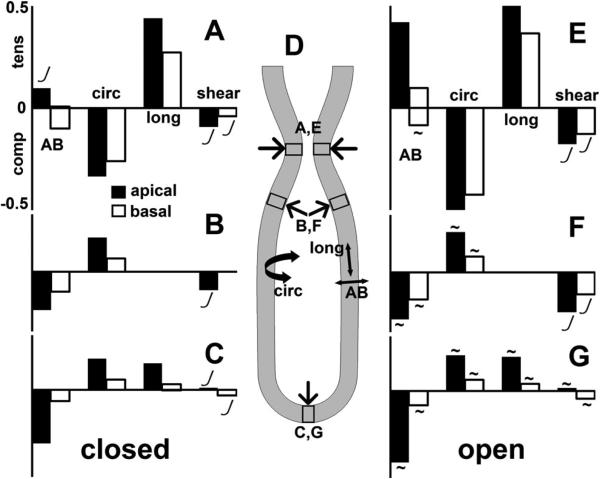
Fig 5.
Stretch (strain) of tissue and cells in the different regions of lung tubule, for closed (A, B, C) and open (E, F, G) trachea, using same inputs. Strains reported in apicobasal (AB), circumferential (circ), and proximodistal/longitudinal (long) directions (D), plus shear. Maximum tensile (tens), compressive (comp), and shear strains reported separately for regions (D) of stenosis (A, E), shoulder (B, F), and tip (C, G). Strains are greater on cells’ apical ends (black bars) than basal ends (white bars). Tensile stretch in one direction is always balanced by compressive stretch in another direction. Strains in the stenotic region (A, E) mainly due to the peristaltic load from the smooth muscle; strains in shoulder (B, F) and tip (C, G) are due to the lumen pressure. Stenotic region (A, E) characterized by circumferential compression up to ~50% balanced by proximodistal extension. Shoulder (B, F) and tip (C, G) regions characterized by apicobasal compression up to ~50%, balanced by extension in the other directions. Larger strains are recorded for open trachea (E, F, G), than for closed trachea (A, B, C). Strains are proportional to force ratio (FT/E), except where marked non-linear (J). Strains are independent of lumen fluid viscosity, except as noted (~).

