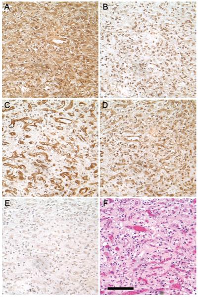
Fig. 1.
Immunohistochemical analysis of notch receptors 1 through 4 in a representative CNS hemangioblastoma. A: NOTCH1 immunoreactivity is distributed uniformly, with cytoplasmic and nuclear staining observed in both the stromal and vascular cells. B: NOTCH2 immunoreactivity is confined to the nucleus. C: NOTCH3 immunoreactivity is strongly expressed in the vascular component of the tumor. D: NOTCH4 immunoreactivity is similar to NOTCH1, with cytoplasmic and nuclear staining observed in both the stromal and vascular cells. E: Nonimmune rabbit IgG control. F: Hematoxylin & eosin staining demonstrates the typical hemangioblastoma appearance with “foamy” stromal cells and abundant vasculature. All antibodies were raised against the C-terminus of the molecule and do not distinguish between the full-length and the cleaved forms. Staining for all four Notch receptors was performed at the same time in the same tumor. Photomicrographs were taken from the same area of the tumor with the same exposure settings, original magnification × 40. Bar = 100 μm.

