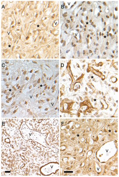
Fig. 2.
Cellular localization of notch receptors. A: NOTCH1 antibody to the C-terminus recognizes both full-length and cleaved NOTCH1 and reacts with both cytoplasmic and nuclear Notch. B: Antibody against NOTCH ICD recognizes only the signaling portion of notch and localizes to the nucleus. The same area of the same tumor is presented in A and B. C: NOTCH2 displays a nuclear localization in stromal cells with only occasional reactivity in vascular cells. D: NOTCH3 staining from the same area presented in panel C reveals an exclusively vascular localization. E: Low-magnification image of NOTCH3 immunoreactivity displaying abundant vasculature. NOTCH3-positive vasculature of multiple morphologies, including large thick- and thin-walled vessels as well as microvessels, is observed. F: NOTCH4 reactivity is seen in the cytoplasm and nuclei of both stromal and vascular cells. Asterisks indicate stromal cells. v = blood vessel. Bar = 20 μm (A–D, and F); bar = 100 μm (E).

