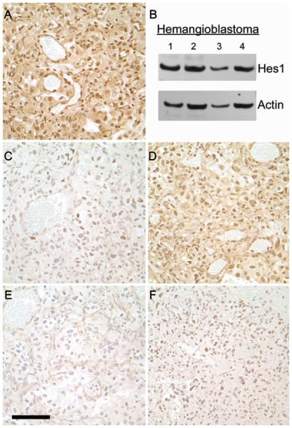
Fig. 3.
Immunohistochemical staining of Notch downstream effectors in a representative CNS hemangioblastoma. A: HES1 was observed in the nuclei of both stromal cells and blood vessels. B: Western blot analysis of 4 hemangioblastomas also demonstrating the presence of HES1. C: Scattered, faint HES3 immunoreactivity observed in the nuclei of some cells. D: HES5 displaying a pattern similar to HES1 although not as intense. E: HEY1 staining is not distinguishable from the negative IgG control (not shown). F: Scattered, faint HEY2 immunoreactivity observed in the nuclei of some cells. Staining for all 5 effector molecules was performed in the same tumor. Original magnification × 40. Bar = 100 μM.

