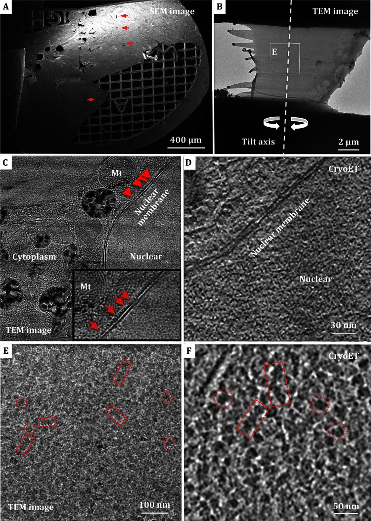
Fig. 4.
Cryo-ultramicrotomy analysis of Hela cells and isolated nuclei. A SEM micrograph of frozen-hydrated Hela cells embedded in ice and attached to Mo-grid with a thin layer of holey carbon supporting film. The lamellas yielded by FIB milling are indicated by red arrows. B Low magnification EM micrograph of the lamella of isolated nuclei. The tilt axis of cryo-electron tomography (cryo-ET) data collection is marked with a dashed line. C TEM image of cryo-FIB milled Hela cells lamella, which shows distinguishable mitochondria (Mt), double nuclear membrane and cytoplasm. The inset image displays the presumptive ribosomes attached to nuclear membrane. D A tomographic slice of cryo-lamella of Hela cells after reconstruction. E Enlarged view of the region highlighted in B with a low dose exposure. F A tomographic slice of the nuclei cryo-lamella after reconstruction. Chromatin-like fibers with longitudinal-section and cross-section orientations are indicated by boxes and circles, respectively, in both E and F

