
Fig. 5.
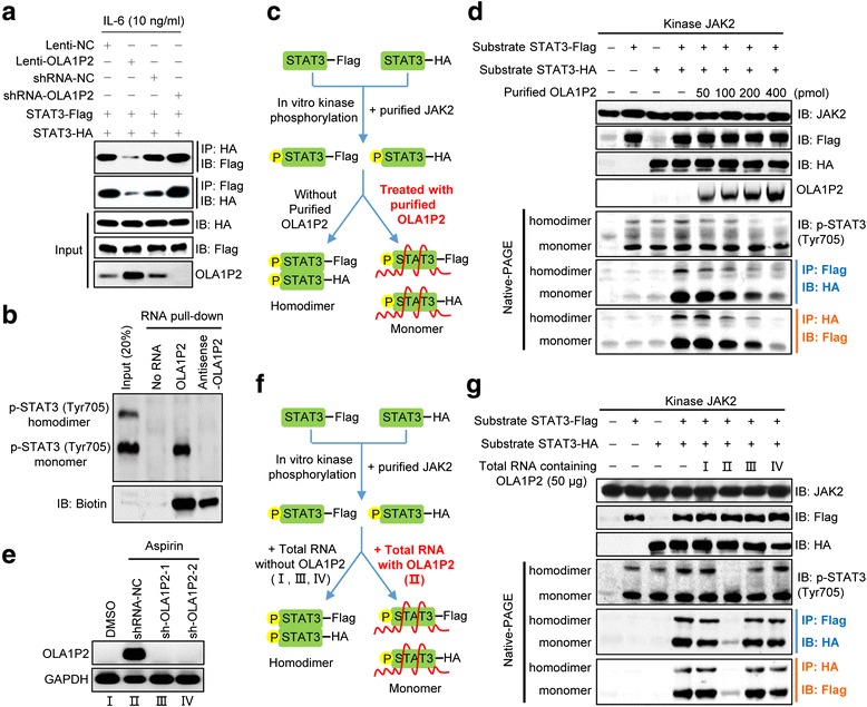
OLA1P2 blocked the formation of phosphorylated STAT3 homodimers. a A total of 293 T cells were transfected with indicated plasmids for 48 h and then treated with IL-6 for 24 h. Immunoprecipitation and immunoblotting analysis were performed to analyze STAT3-Flag and STAT3-HA protein levels. b COLO205 cells were subjected to RNA pull-down analysis using 5′ biotin-linked RNAs, and the eluted proteins were determined using immunoblotting analysis following polyacrylamide gel electrophoresis under non-denaturing conditions. c Schematic diagram showing the experimental design of purified OLA1P2 blocking the formation of phosphorylated STAT3 homodimers. We used 4 μg purified STAT3 (2 μg STAT3-Flag combined with 2 μg STAT3-HA) incubated with 2 μg purified JAK2. After incubation, purified OLA1P2 was then added to the reaction mixture. d Different amount of purified OLA1P2 blocking the formation of phosphorylated STAT3 homodimers. e QRT-PCR analysis of the amount of lncRNA OLA1P2 in COLO205 cells treated with aspirin and shRNA-OLA1P2. f Schematic diagram showing the modified experimental design of total RNA containing OLA1P2 blocking the formation of phosphorylated STAT3 homodimers. We used 4 μg purified STAT3 (2 μg STAT3-Flag combined with 2 μg STAT3-HA) incubated with 2 μg purified JAK2. After incubation, total RNA extracted from COLO205 cells were then added to the reaction mixture. g The total RNA of OLA1P2 silencing COLO205 cells failed to block the formation of phosphorylated STAT3 homodimers