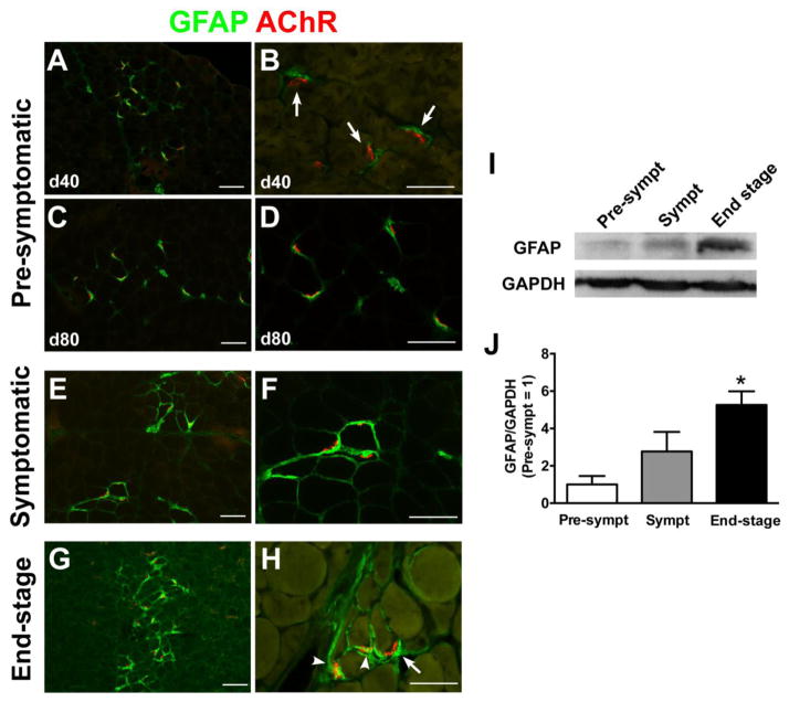
Figure 2. GFAP expression is increased in the muscle of SOD1G93A rats following disease progression.
(A–H) GFAP immunostaining in the hindlimb muscle of SOD1G93A rats. GFAP expression was limited near AChR-positive endplates at 40 days (A, B) and 80 days (C, D) of age. However, GFAP expression gradually increased in symptomatic (E, F) and end stage (G, H). (I) Representative band images of western blotting for GFAP and GAPDH expression. (J) Densitometric analyses of western blotting data supported that GFAP was increased in the muscle homogenates of end-stage rats. Scale bar: 100 μm in A, C, E, G; 50 μm in B, D, F, H. *: P<0.05 vs. pre-symptomatic and symptomatic.

