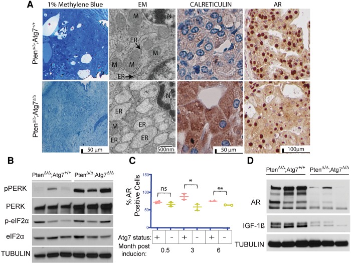
Figure 4.
Autophagy deficiency results in ER stress and decreased AR signaling. (A) One percent methylene blue-stained tissue showing representative areas used for EM analysis. Representative transmission EM (TEM) with ER and mitochondria (M) labeled as well as calreticulin and AR IHC of Atg7+/+ and Atg7Δ/Δ anterior mouse prostates at 3 mo after tumor induction. (B) Western blot analysis of pPERK, PERK, p-eIF2α, and eIF2α expression in tumor lysates from Atg7+/+ and Atg7Δ/Δ anterior mouse prostates at 3 mo after tumor induction. Tubulin served as a loading control. (C) Quantification of the percentage of cells with AR-positive nuclei in Atg7+/+ and Atg7Δ/Δ anterior mouse prostate tissue (n = 2) at various times after tumor induction. The Atg7+/+ tumors had 88.6% and 81.35% AR-positive nuclei at 3 and 6 mo, respectively, whereas the Atg7Δ/Δ tumors had only 58.2% and 65.45% positive nuclei at 3 and 6 mo after TAM, respectively. (D) Western blot analysis of AR and IGF-1β expression in Atg7+/+ and Atg7Δ/Δ mouse prostates at 3 mo after tumor induction.

