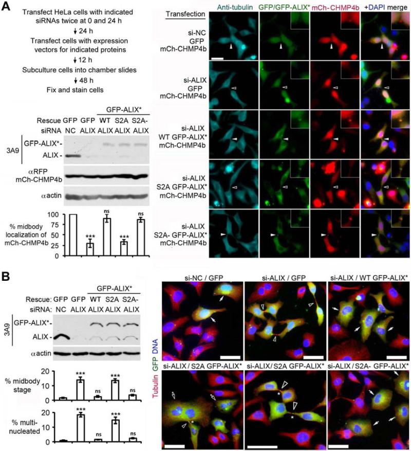
Figure 5. The S718-S721 phosphorylation is required for ALIX to function in cytokinetic abscission.
(A) HeLa cells were transfected with the indicated agents and cultured as diagrammed, followed by IB of cell lysates with the indicated antibodies. Fixed cells were stained with αtubulin and αGFP, and GFP-mCherry double positive cells were scored for the midbody localization of mCh-CHMP4b (n = 3 ± SD). Solid and hollow arrowheads in the representative images indicate the presence or absence of mCh-CHMP4b at the midbody, respectively. Enlargements (3×) depict the midbody area; scale bar: 50 μm. (B) HeLa cells were transfected with the indicated agents and cultured as done in (A), followed by IB of cell lysates with the indicated antibodies. Fixed cells were stained with αtubulin and αGFP, and GFP positive cells were scored for midbody-stage or multinucleation (n = 3 ± SD). Solid and hollow arrows in representative images indicate GFP positive mononucleated and multinucleated cells, respectively; hollow arrowheads indicate midbodies between GFP positive cells; and the asterisks indicate the midbody localization of GFP-ALIX*. Scale bar: 50 μm. See also Figure S3.

