Fig. 4.
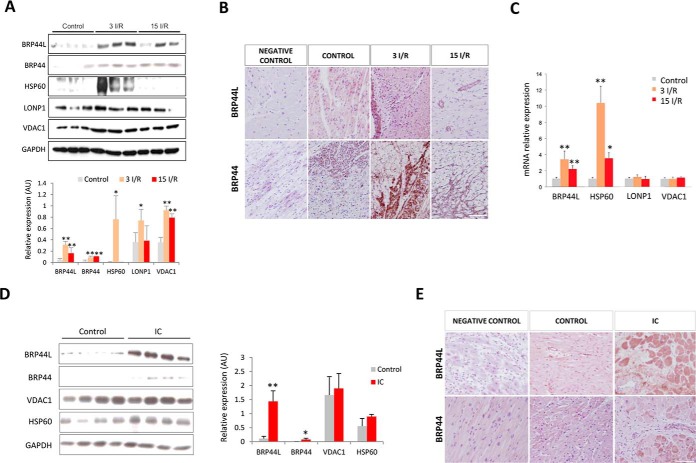
Validation of the proteomic data by alternative techniques in the porcine model and in heart tissue from ischemic cardiomyopathy (IC) patients. A, Western blot and quantitative densitometry confirming overexpression of BRP44L, BRP44, HSP60, LONP1, and VDAC1 in the border zone from porcine heart samples. The protein normalized value is indicated for each sample. B, immunohistochemistry of paraffin-embedded pig heart tissue in the healthy control group, close to the infarcted area, 3 (3 I/R) and 15 days (15 I/R) after I/R. C, BRP44L, HSP60, LONP1, and VDAC1 mRNA levels in the same porcine zones. D, Western blot analysis and quantitative densitometry of BRP44L, BRP44, VDAC1, and HSP60 expression in heart tissue from healthy donors (Control) and patients undergoing cardiac heart transplantation. The normalized protein value is indicated for each sample. E, immunohistochemistry of paraffin-embedded healthy and human heart tissue close to the infarcted area. Scale bar, 50 μm; * indicates p value <0.05; ** indicates p value <0.001.

