
Figure 6.
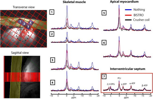
The 3D‐CSI cardiac spectra acquired with three suppression protocols: no saturation (blue spectra); BISTRO saturation (red spectra); and using the crusher coil with Ispoil × Tspoil = 0.9 A·ms (black spectra) (experiment 4). Left: CSI matrix overlaid on 7T CINE FLASH localizers showing the locations of voxels 1–7 and of the BISTRO saturation band (in yellow). Voxels are shown that contain primarily skeletal muscle (blue) or myocardium (red). Voxel 7 is representative of the interventricular septum and was quantified with AMARES as shown in Table 2. The dashed circle denotes the 50% of maximum point spread function contour for voxel 7 (i.e., the “true” voxel size).