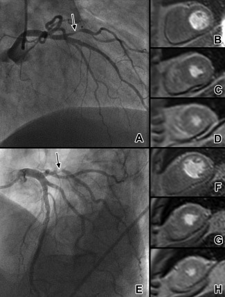
Figure 3.

Example of two typical patients with false‐negative MR perfusion. A patient with diagonal branch stenosis (A, arrowed) and absence of a clear perfusion defect on basal (B), mid‐ventricular (C), and apical (D) first‐pass perfusion MR images. SPECT imaging showed a subtle anterolateral perfusion defect. Duke jeopardy score was 2. Another patient with obtuse marginal branch stenosis (E, arrowed) again has no visible perfusion defect on MR (F–H). SPECT was negative in this case, with Duke jeopardy score of 2. MR appears to more commonly miss stenoses which subtend relatively small areas of myocardium.
