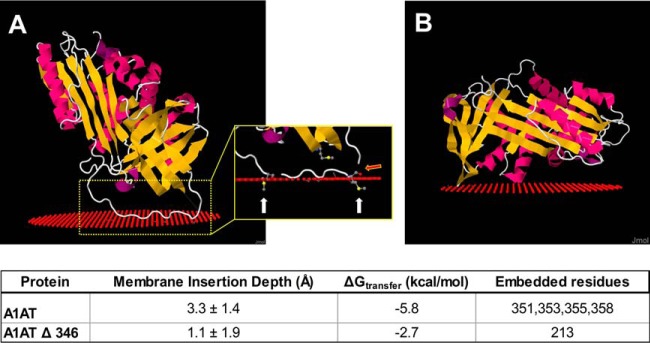
Fig. 6.
Structural prediction of lipid binding by α-1-antitrypsin. A, Predicted binding of α-1-antitrypsin (A1AT) to a lipid surface (red spheres). Inset demonstrates that methionine residues (Met351 and Met358) are embedded in the lipid (white arrows) and indicates the cut site for neutrophil elastase (red arrow). B, Predicted lipid binding of A1AT structure with the reactive center loop removed (A1AT Δ 346).

