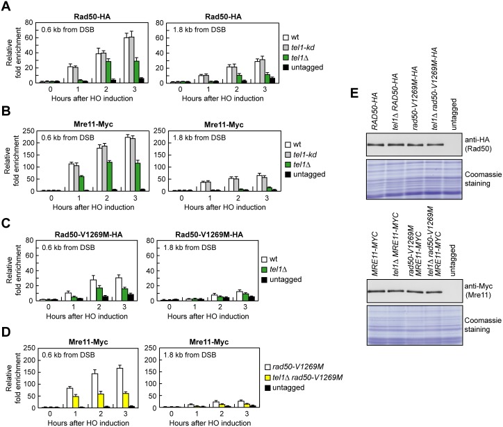
Fig 7. The lack of Tel1 impairs MRX and MRV1269MX association to a DSB.
(A–D) ChIP analysis. Exponentially growing YEPR cell cultures were transferred to YEPRG at time zero. Relative fold enrichment of the indicated fusion proteins at the indicated distances from the HO cleavage site was determined after ChIP with anti-HA or anti-Myc antibodies and subsequent qPCR analysis. Plotted values are the mean values with error bars denoting s.d. (n = 3). (E) Western blot with anti-HA or anti-Myc antibodies of extracts used for the ChIP analysis shown in (A–D). The same amount of protein extracts was separated on a SDS-PAGE and stained with Coomassie Blue (loading control).

