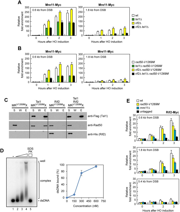
Fig 8. Rif2 is recruited to DNA ends, and its lack enhances MRX and MRV1269MX association to the DSB.
(A, B) ChIP analysis. Exponentially growing YEPR cell cultures were transferred to YEPRG at time zero. Relative fold enrichment of Mre11-Myc fusion protein at the indicated distances from the HO cleavage site was determined after ChIP with anti-Myc antibodies and subsequent qPCR analysis. Plotted values are the mean values with error bars denoting s.d. (n = 3). (C) Flag-tagged Tel1 (50 ng) was incubated with MRV1269MX (100 ng) in the absence or presence of Rif2 (100 ng), and protein complexes were captured by anti-Flag resin and followed by immunoblotting analysis. (D) Rif2 (150, 300 and 600 nM) was incubated with 32P-labeled 100-bp dsDNA (10 nM) in the presence of ATP. In lane 5, the reaction mixture was deproteinized with SDS and proteinase K (PK) prior to analysis. Plotted values are the mean value with error bars denoting s.d. (n = 3). (E) ChIP analysis. As in (A), but showing Rif2 recruitment at the HO-induced DSB.

