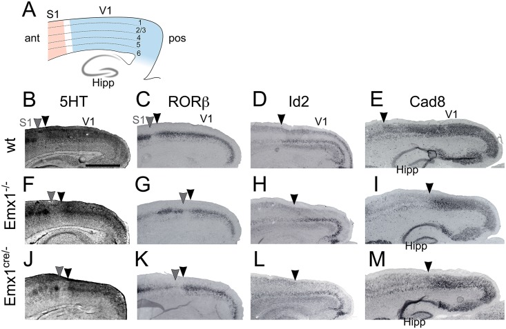
Fig 1. Gene expression demonstrates a reduction in V1 size with normal laminar expression following Emx1 deletion.
Immunostaining and in situ hybridization for several markers was performed on sagittal sections of P7 cortices. (A) Schematic of a sagittal section detailing the cortical layers and location of the hippocampus (Hipp). Stained sections of wt (B–E), Emx1-/- (F–I), and Emx1cre/- (J–M) have been cropped to highlight changes in V1. (B, F, J) 5HT immunostaining revealed layer 4 of V1 and S1. Compared to wt, Emx1 deletions (Emx1-/- and Emx1cre/-) possessed a smaller, posteriorly shifted V1 (black arrowheads denote anterior edge of V1, gray arrowheads denote posterior edge of S1). (C, G, K) RORβ is expressed in layer 4 of primary sensory areas. In Emx1 deletions the posterior shift of the anterior edge of V1 (black arrowheads) and posterior edge of S1 (gray arrowheads) can be appreciated, showing the reduction in V1 size. (D, H, L) Id2 is expressed in layer 5 of V1 and also showed a posterior shift of the anterior edge of V1 in Emx1 deletions (arrowheads). (E, I, M) Cad8 is expressed in layers 2/3 of V1, and in Emx1 deletions the anterior edge of V1 (arrowheads) was shifted posteriorly, and V1 size was reduced. Cad8 is also expressed in the hippocampus, which is ventral to the cortex and provides a stable landmark for comparison between genotypes. All of the stains presented demonstrated the reduction of V1 in the sagittal plane, as well as the posterior shift of V1. However all of these markers were expressed in the appropriate cortical layers. Anterior is to the left, dorsal to the top. V1, primary visual area; S1, primary somatosensory area; Hipp, hippocampus; ant, anterior; pos, posterior. Scale bar, 1.0 mm. Bar in B applies to all panels.

