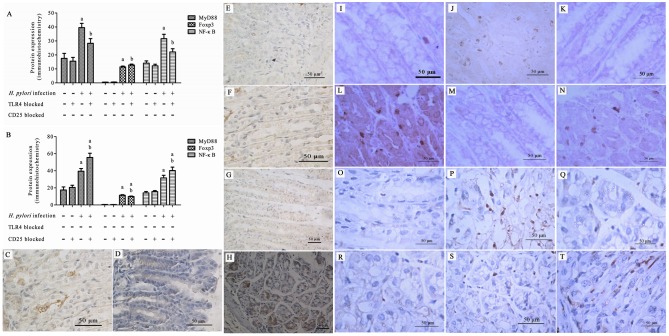
Fig 4. Expression of MyD88, NF-κB p65, and Foxp3 in the gastric mucosa by immunohistochemistry after H. pylori infection.
Expression of MyD88, NF-κB p65, and Foxp3 with TLR4 (A) or CD25 (B) blocked; (C-T) Representative images of immunohistochemical staining. MyD88 staining in the gastric mucosa of the untreated (C, D), TLR4 blocked (E, F), or CD25 blocked (G, H) control and H. pylori infection groups; NF-κB p65 staining of the gastric mucosa of the untreated (I, J), TLR4 blocked (K, L), or CD25 blocked (M, N) control and H. pylori infection groups; Foxp3 staining of the gastric mucosa of the untreated (O, P), TLR4 blocked (Q, R), or CD25 blocked (S, T) control and H. pylori infection groups. aP < 0.001 vs. control or TLR4 blocked control groups; bP < 0.001–0.05 vs. control and TLR4 blocked control groups and the H. pylori group (Fig 4A). aP < 0.001–0.01 vs. control and CD25 blocked control groups; bP < 0.01–0.05 between CD25 blocked H. pylori and H. pylori groups (Fig 4B).

