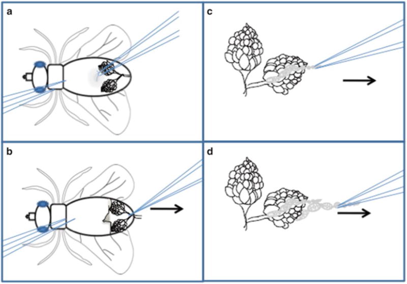
Fig. 2.

Ovary dissection technique: Schematic representation of egg chamber dissection. (a) The female fly should be immobilized on its back using the left forceps. The right forceps are used to pinch the soft cuticle of the ventral side of the abdomen. (b) Pull the cuticle toward the right (arrow), revealing the ovaries. Grasp the common oviduct with the right forceps and pull to the right (arrow) to free the ovaries from the carcass. (c) Immobilize the ovary pair by pinching the oviduct using the left forceps. Use the right forceps to grasp the tip of an ovariole and slowly pull to the right (arrow). (d) Repeat until multiple ovarioles have been freed from the sheath
