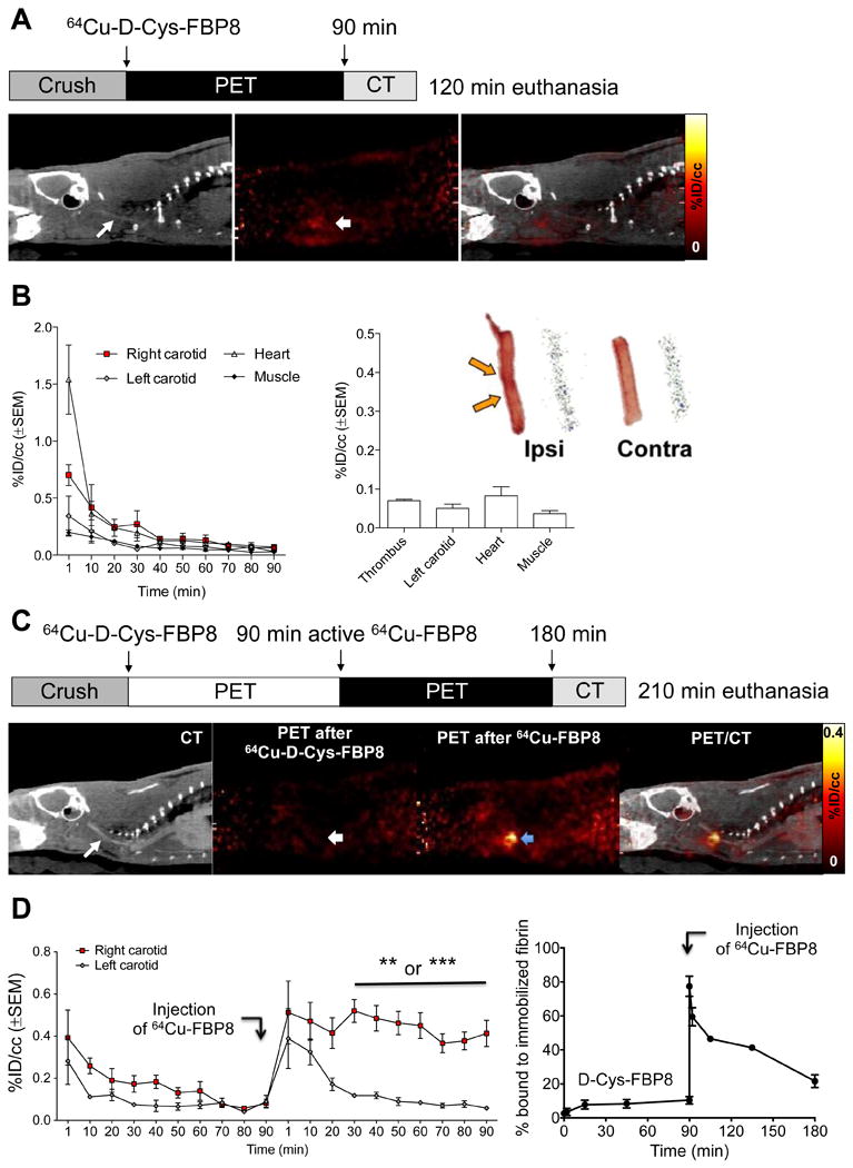
Figure 4.

(A) CT, PET, and fused images from an animal injected with 64Cu-D-Cys-FBP8 after carotid crush injury (n = 2). Contrast-enhanced CT angiography was used to detect the common carotid artery (thin arrow). The thrombus location could not be detected by PET. (B) Time–radioactivity curves obtained from PET imaging, mean radioactivity values (30 - 90 min p.i.), and representative photograph and autoradiograph of ipsilateral and contralateral carotid arteries showing comparable radioactivity levels between thrombus and background. (C) PET-CT imaging (n = 3) after sequential injection of 64Cu-D-Cys-FBP8, followed by injection of its active analogue 64Cu-FBP8. Thrombus uptake was only detected after injection of 64Cu-FBP8 (blue arrow). (D) Statistically significant difference in thrombus versus contralateral artery uptake for 64Cu-FBP8 but not 64Cu-D-Cys-FBP8. In vitro binding studies of blood plasma incubated with immobilized fibrin showed significantly lower binding for 64Cu-D-Cys-FBP8 in comparison to 64Cu-FBP8 (n = 2). **P <0.01.
