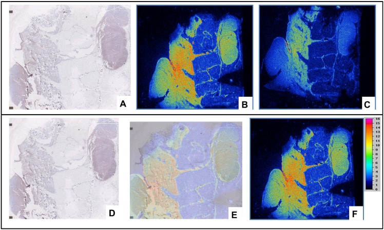
Fig 8.
(A) Immunostaining of nodular melanoma using anti-hMMP-9 murine monoclonal antibody ab58803. Radiolabeling of adjacent tissue sections of nodular melanoma with 111In-F3B-DOTA (B), and 111In-DOTA-control sequence (C). Comparison of the results obtained by radiolabeling with 111In-DOTA-F3B and by immunostaining. The results of immunohistochemical images corresponded with area with high activity. (D) Immunostaining of nodular melanoma with ab58803 murin monoclonal antibody anti-human MMP-9. It’s possible to identify the primitive nodular melanoma and an intrahypodermic node (E) Merged Images. (F) Radiolabeling with 111In-DOTA-F3B.

