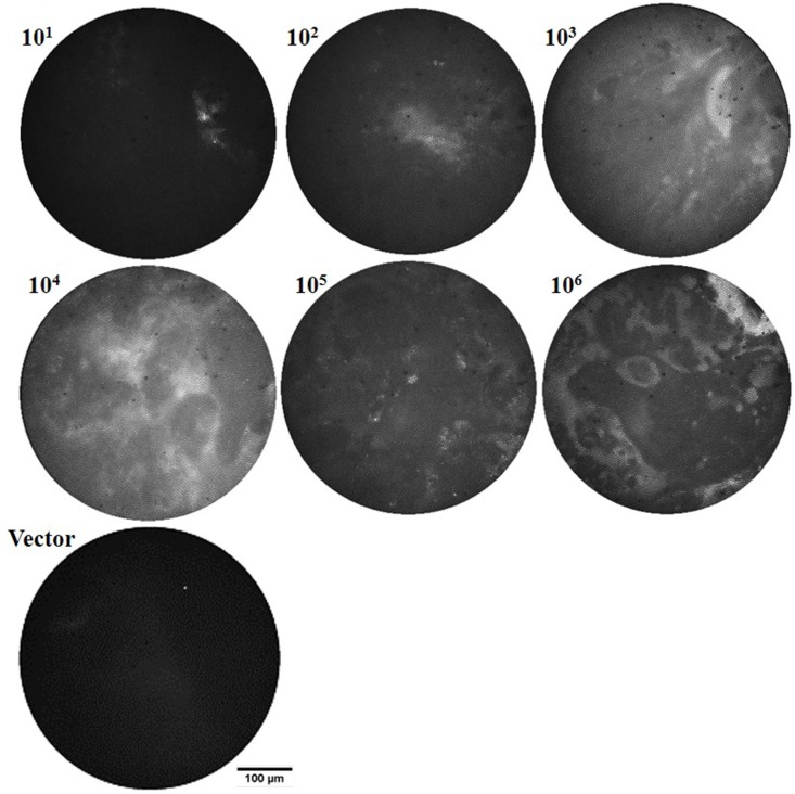
Fig 5. In vivo microendoscope images provide feedback for positioning the intravital excitation source and can allow a qualitative evaluation of bacterial loads in the lungs.
Representative in vivo fiber microendoscopy images of infected mice lungs, 24 hr post-infection with 101−106 colony forming units (CFU) BCG17 (tdTomato expressing BCG strain) and 105 CFU BCG39 (BCG containing the same vector that does not express tdTomato (Vector)).

