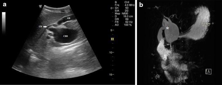
Fig. 1.
Combined dilatation of both the cystic duct as well as the common bile duct in a 14-year-old boy who presented with mild epigastric discomfort. a Abdominal ultrasound image showing a cystic lesion involving the common bile duct (CBD) and the adjacent portion of the cystic duct (CD), GB (gall bladder); b thick-slab coronal oblique MR cholangiopancreatography image showing fusiform dilatation of the CBD involving the cystic duct via a wide communication (asterisk)

