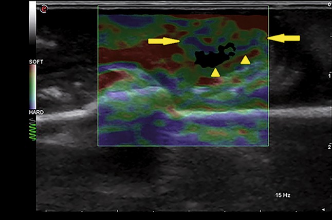
Fig. 5.

Elastography scan performed on the adequately prepared surgical specimen. The tumor (arrows and arrowheads) is represented by a mosaic of colors (mainly green, turquoise and blue) suggestive of a greater stiffness than the adjacent wall (mainly red)
