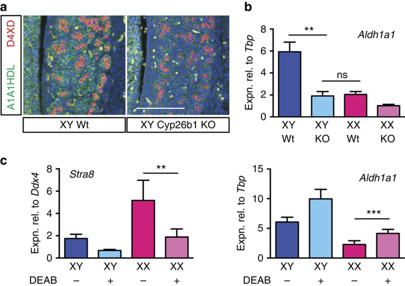
Figure 5. ALDH1A1 expression responds to levels of RA present.
(a,b) Immunofluorescence (a) and qRT–PCR (b) analyses show ALDH1A1/Aldh1a1 expression in 13.5 d.p.c. fetal testis is diminished when Cyp26b1 is deleted (for b, n=3, 5, 3, 2), scale bar, 100 μm. (c) When 11.5 d.p.c. UGRs are cultured in the presence of DEAB (ALDH inhibitor) for 48 h Stra8 expression is diminished, as expected, but Aldh1a1 expression is augmented in ovary suggesting that in Aldh1a2/3-knockout ovaries Aldh1a1 expression may be elevated above normal levels (n=2, 2, 5, 7). NS, not statistically significant, **P<0.01, ***P<0.001 (Student's t-test, unpaired for b, paired for c, mean+s.e.m. shown).

