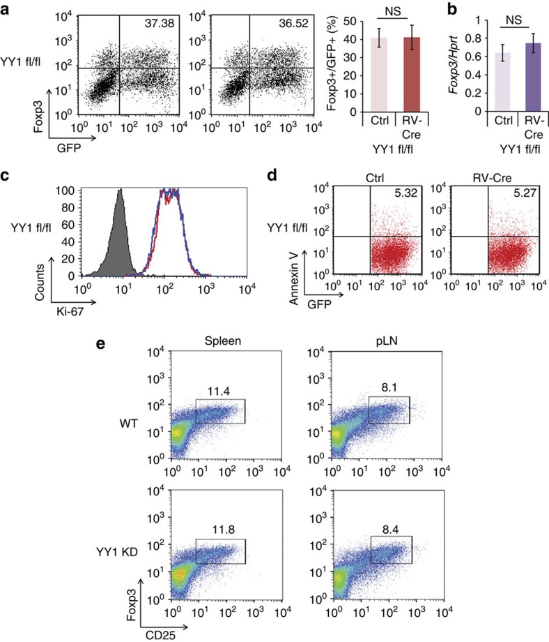
Figure 3. YY1 deficiency does not influence Foxp3 expression.
(a) Naïve CD4 T cells from YY1 fl/fl mice were transduced with either control vector or CRE-expressing vector (RV-Cre) and cultured under the Treg differentiation condition for 4 days. Foxp3 was stained and measured by flow cytometry. Numbers in the FACS plots indicate the percentage of Foxp3+ cells from GFP+ cells. (b) GFP+ cells from a were sorted, and total RNA was isolated. A relative amount of Foxp3 was measured by qRT–PCR. (c) Proliferation of YY1-deficient Treg cells was detected with Ki-67 staining from GFP+ cells. (d) Apoptosis of YY1-deficient Treg cells was measured with Annexin V staining. Experiments were performed five times with similar results. (e) Treg populations in the spleen and peripheral lymph nodes of YY1 KD or WT mice were analysed by staining anti-CD4, anti-CD25 and anti-Foxp3. Cells were gated on CD4.

