Figure 6.
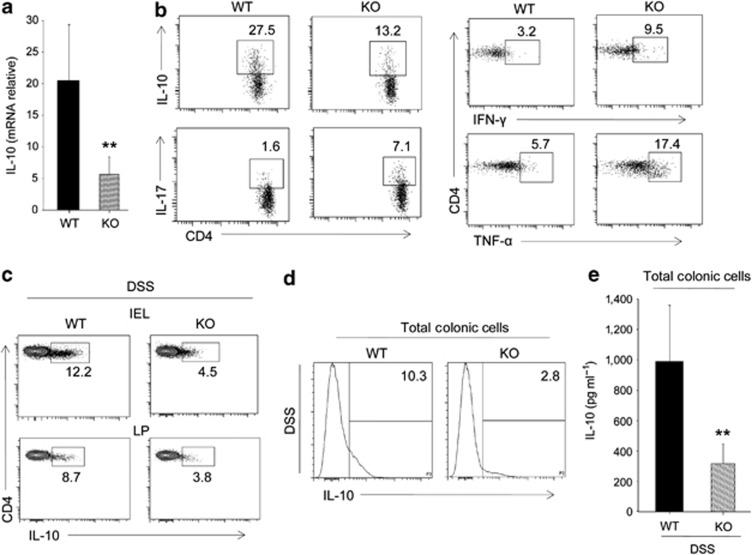
SMAR1-deficient transforming growth factor (TGF)-β1-induced Treg cells showed reduced expression of interleukin (IL)-10 and elevated levels of proinflammatory cytokine genes. (a) Amount of IL-10 mRNA in TGF-β1 induced Treg cells from wild-type (WT) and SMAR1−/− mice evaluated by quantitative real-time PCR (**P<0.001, Student's t-test). (b) Flow cytometry of intracellular production of IL-10, IL-17, interferon (IFN)-γ, and tumor necrosis factor (TNF)-α in TGF-β1-induced Treg cells from WT and SMAR1−/− mice. The numbers in the plot indicate the percentage of CD4+ T cells. Data are representative of three independent experiments with similar results. (c,d) Flow cytometry analysis of intracellular IL-10 in CD4+ T cells (c) and total colonic cells (d) isolated from colon lamina propria (LP) and epithelium (intraepithelial lymphocyte (IEL)) at seventh day of dextran sodium sulfate (DSS)-treated WT and SMAR1−/− mice. The numbers in the plots indicate the percentage of cells in each; data are representative of three independent experiments with six mice per group. (e) Enzyme-linked immunosorbent assay analysis of IL-10 levels in total colonic LP cells at seventh day of DSS-treated WT and SMAR1−/− mice (mean±s.d. six mice per group). Data represent three independent experiments (**P<0.002, Student's t-test). KO, knock out.

