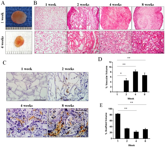
Fig 3. Vascular infiltration in collagen scaffolds implanted in mice.
(A) Appearance of scaffolds harvested from mice at 1 and 4 weeks. Representative images of scaffold sections stained with haematoxylin and eosin (B) or endothelial cell marker CD31 (C) at 1, 2, 4 and 8 weeks post-implantation. Percentage of vascular volume (D) and percent of remaining collagen scaffold (E) at 1, 2, 4 and 8 weeks post-implantation (n = 3–6 mice). *p<0.05 and **p<0.01 by one-way ANOVA with Turkey post-hoc test. Scale bar = 100 μm.

