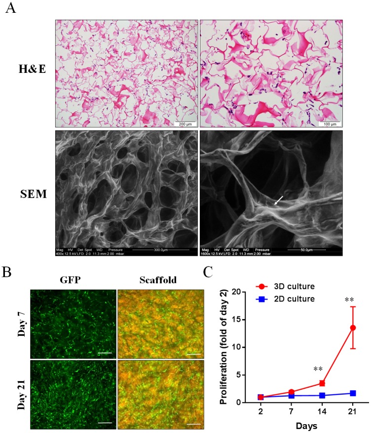
Fig 5. Morphology and growth of human ASCs cultured in 3D collagen scaffolds over 21 days.

(A) Representative images captured from 3D collagen scaffolds seeded with human ASCs for 24 hours: haematoxylin and eosin (H&E) staining and scanning electron microscopy (SEM). The white arrow demonstrates human ASCs attached to the collagen scaffold. (B) Representative images of GFP-expressed human ASCs cultured in collagen scaffolds for 7 and 21 days. Scale bar = 200 μm. (C) The proliferation rate of GFP-expressed human ASCs cultured in 3D collagen scaffolds and on 2D collagen-coated plates (n = 3). **p<0.01 vs 2D culture by unpaired t-test.
