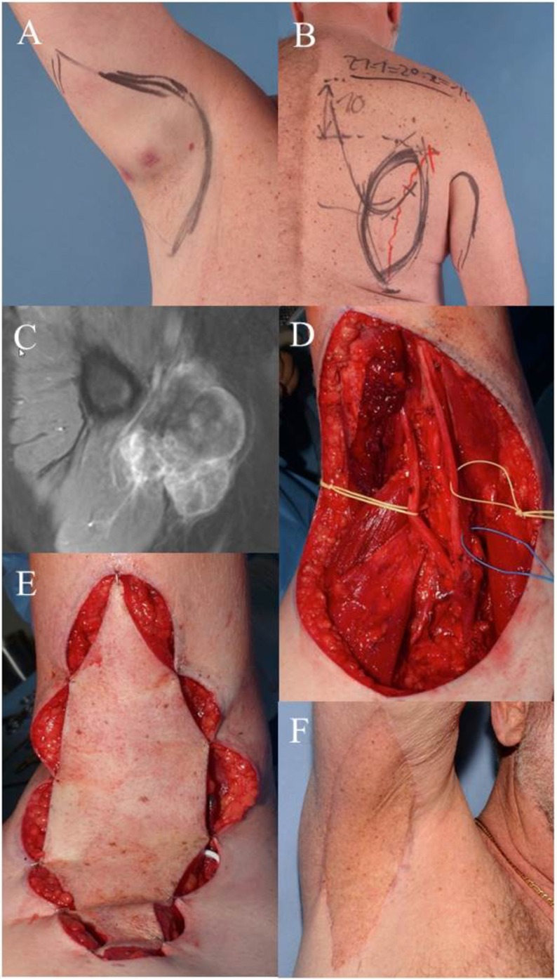
Figure 1.
Case 1: (A) clinical visible mass in the right axilla, pre-OP drawing of the wide resection margins. (B) Pre-OP drawing demonstrates planned pedicled parascapular fasciocutaneous flap. (C) Pre-OP MRI of the right axilla. (D) Intraoperative situation after wide en-bloc excision of tumor. Deep margins were achieved by epineurectomy and adventitiectomy. (E) Pedicled parascapular flap inset reconstructs the defect. (F) Clinical appearance 5 years after surgery and external post-OP radiation therapy.

