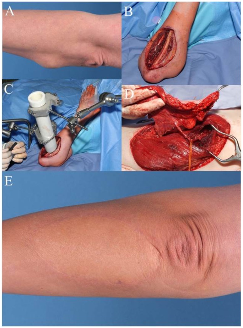
Figure 2.
Case 2: (A) pre-OP picture shows mass in the proximal forearm. (B) Intra-OP situation after wide en-bloc excision. (C) Intra-OP application of radiation therapy in the wound bed close to the ulna (15 Gy). (D) Raised anterolateral thigh flap from the left leg for microvascular reconstruction. (E) Clinical appearance of the reconstructed forearm 1 year after additional external radiation therapy (50.4 Gy).

