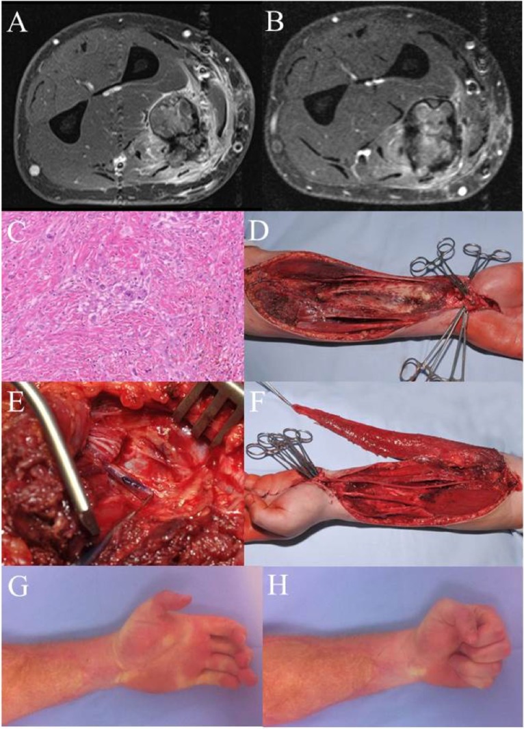
Figure 3.
Case 3: (A) MRI left forearm before radiation. (B) MRI left forearm post radiation. (C) H&E specimen showing only few scattered remaining tumor cells as a result of the preoperative radiotherapy. (D) Intraoperative situation after en-bloc excision of the flexor compartment. (E) Intraoperative detail photography of the transected median nerve. The blue marking shows the motor branch to the deep flexors, later used for nerve coaptation to the obturator branch of the transferred gracilis muscle. (F) Gracilis muscle after microvascular transfer and motor nerve coaptation. After defining the proper tension, the muscle will be fixed to the deep flexor tendons (II–V). Flexor pollicis longus function was reconstructed using a brachioradial tendon transfer. (G,H) Clinical result 5 years after therapy. There is a remaining extension deficit, but a full finger flexion with a strong grip could be achieved. Patient is in complete remission and rehabilitated in his original job as a truck driver.

