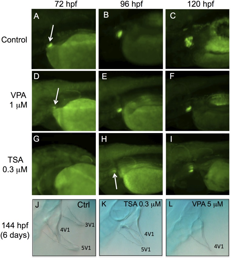
Fig 6. VPA and TSA delay the formation of pharyngeal teeth.
The development of pharyngeal teeth was analyzed at 72 hpf, 96 hpf and 120 hpf. GFP fluorescence in embryos of the Dlx2b-EGFP transgenic line: controls (A-C), embryos treated with 1 μM VPA (D-F) or 0.3 μM TSA (G-I). White arrows indicate pharyngeal tooth germs. Alcian blue staining at 144 hpf in control embryos (J) or embryos treated with 0.3 μM of TSA (K) or 5 μM of VPA (L). Dotted lines highlight the contours of three teeth: 3V1, 4V1 and 5V1.

