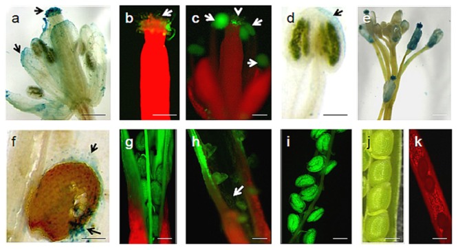
FIGURE 3.

U. virens-Arabidopsis flower interaction. (a) Visible intense GUS histochemical staining on different floral regions of infected Arabidopsis flower by U. virens 3 weeks after post-inoculation; (b) GFP tagged U. virens hyphae growing on the stigma 3 weeks after post-inoculation; (c) GFP-tagged aerial mycelia on the male and female parts of Arabidopsis flower 3 weeks after post-inoculation; (d) U. virens infected male floral structure showing the GUS stain; (e) Colonization of GUS labeled aerial mycelia on the stem, anther, and stigma of the flowers after 3 weeks of post inoculation; (f–i) Mycelium development on siliques and seeds as well as shriveled pod formation from infection of flower tissue 28 days after inoculation with GFP and GUS labeled and transformed strains of U. virens; (j) Control showing Arabidopsis siliques and seeds; (k) Uninfected Arabidopsis pod under epifluorescence microscopy. Bars = 40 μm (g–j), 20 μm (a–d,f,k), 150 μm (e).
