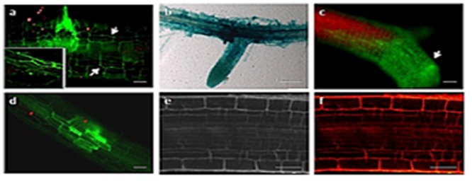
FIGURE 4.

Early colonization stages of Arabidopsis roots by U. virens. (a) hyphae growing along the epidermis parallel to the longitudinal axis of the root and forming lateral hyphopodia (arrow), 2 dpi. The inset shows magnification of the upper section of the root; (b) younger GUS stained hyphae extensively colonize the root surface, forming a network around the root; (c) heavy colonization of the Arabidopsis root by U. virens after 10 dpi; (d) root cells become colonized by hyphae in a mosaic pattern, leaving some cells uninfected after 10 dpi; (e) root of the uninfected Arabidopsis plant under bright field microscopy (Control); (f) uninfected Arabidopsis root under epifluorescence microscopy. Bars = 100 μm (a,c,d), 40 μm (e,f) and 50 μm (b).
