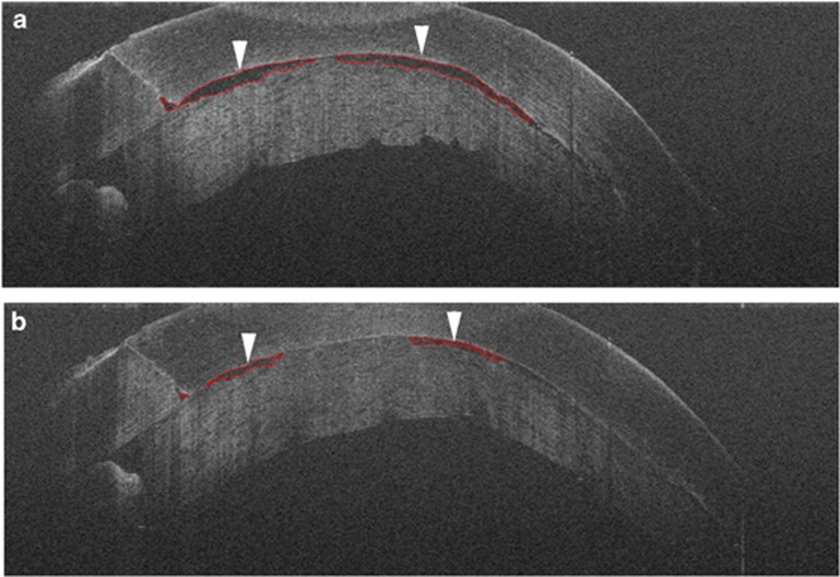
Figure 2.
Intraoperative OCT for Descemet stripping automated endothelial keratoplasty with automated interface fluid analysis. (a) Time point 1 shows interface fluid between the graft and host (arrowheads) with automatic segmentation around fluid to evaluate overall fluid metrics (red lines). (b) Time point 2 shows reduction of interface fluid (arrowheads) following corneal manipulation with successful automated identification of fluid interface (red lines).

