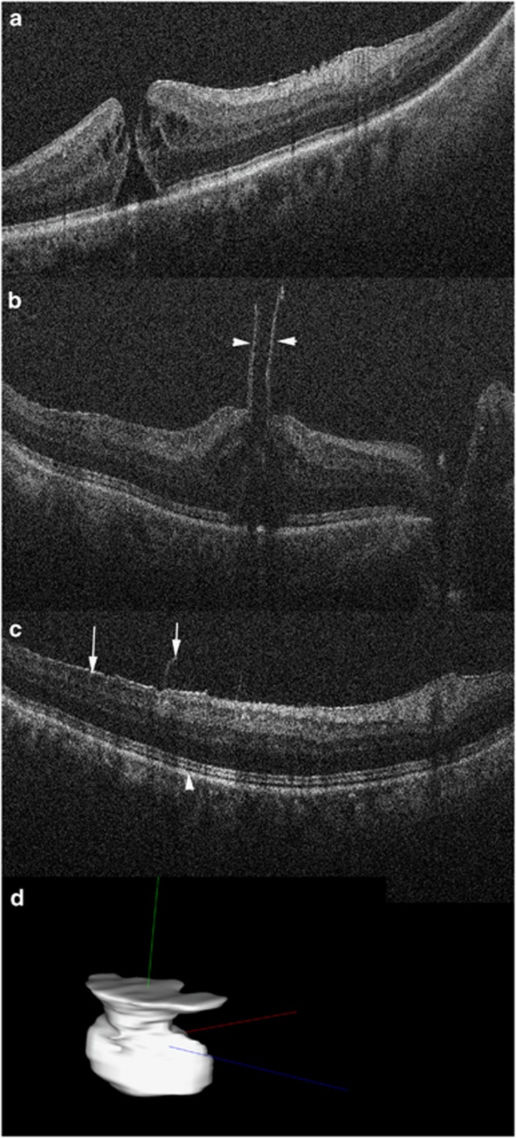
Figure 3.
Intraoperative OCT for macular hole surgery. (a) Preincision OCT revealing full-thickness macular hole with minimal associated epiretinal membrane. (b) Post-peel OCT confirms large residual flap at edge of hole (arrowheads). Configuration information may be used to guide inverse internal limiting membrane flap technique. (c) Post-peel OCT also identified more peripheral residual epiretinal membrane edges (arrow) and expansion of the ellipsoid zone to retinal pigment epithelium height (arrowhead, compare with A). (d) Automated volumetric segmentation of macular hole with three-dimensional reconstruction allowing for visualization of alterations between pre- and post-peel configurations.

