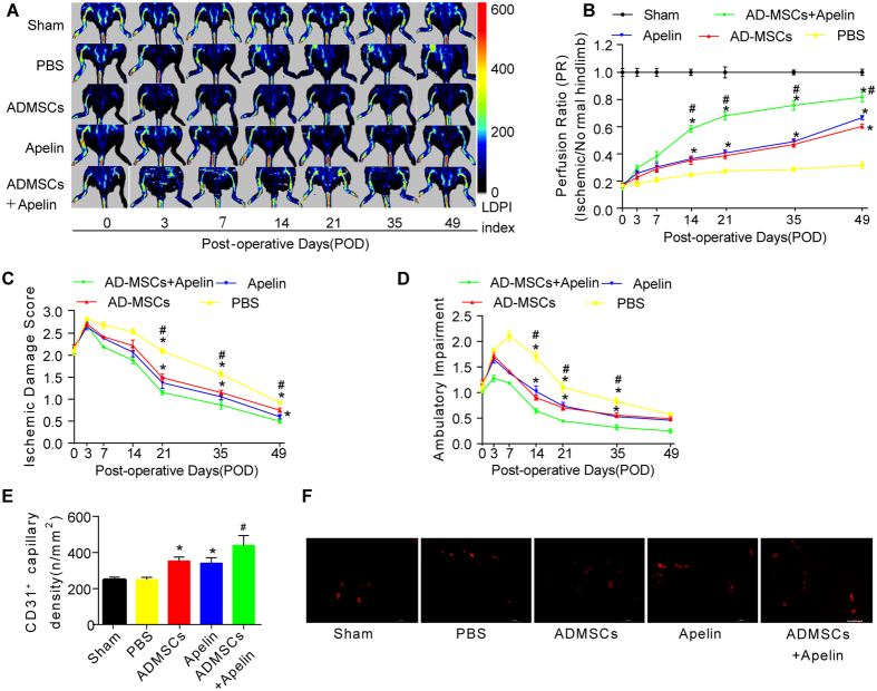
Figure 2. AD-MSCs transplantation and apelin administration promoted hindlimb functional recovery and angiogenesis in the PAD model.
(A,B) In vivo laser Doppler perfusion imaging (LDPI) visualized dynamic changes in hindlimb blood perfusion, which was (B) quantified using perfusion ratio (PR), i.e., the ratio of average LDPI index of ischemic (left leg, red arrows, the same to fig. 4A) to nonischemic hindlimbs. Colored scale bar represents blood flow velocity in LDPI index. n = 10. (C,D) Cumulative results for functional assessment of ischemic muscle over follow-up are shown graphically as ischemic damage score (C) and ambulatory impairment score (D), n = 15 for each group. (E,F) Representative image and quantitative analysis of the CD31-positive blood vessels within the same-sized regions of adductor muscle section among groups, as assessed by immunofluorescence staining with endothelial marker CD31 (PECAM-1) on POD49. n = 20 random fields. Error bars represent mean ± SD. *P < 0.05 vs. PBS, #P < 0.05 vs. both AD-MSCs and apelin. Scale bar: 100 μm.

