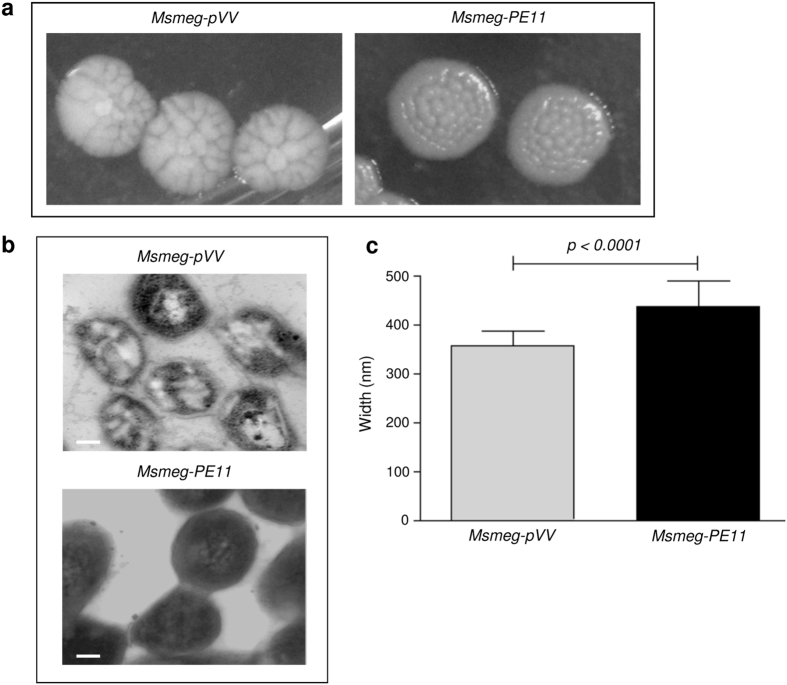
Figure 1. PE11 alters colony morphology, surface architecture and width of M. smegmatis.
(a) Msmeg-pVV or Msmeg-PE11 bacteria were plated on 7H10 agar plates supplemented with OADC and incubated for 5–6 days and the mycobacterial colonies were photographed. (b) For transmission electron microscopy (TEM) analysis, Msmeg-pVV or Msmeg-PE11 strains were cultured on 7H10 agar for 5–6 days and surface architecture of these bacteria were analyzed. Scale bar, 100 nm. (c) In another experiment, Msmeg-pVV or Msmeg-PE11 bacteria were harvested for scanning electron microscopy (SEM) analysis to measure the diameters of Msmeg-pVV or Msmeg-PE11 and mean width of 100 bacilli each of Msmeg-pVV and Msmeg-PE11 is graphically represented in nm.

