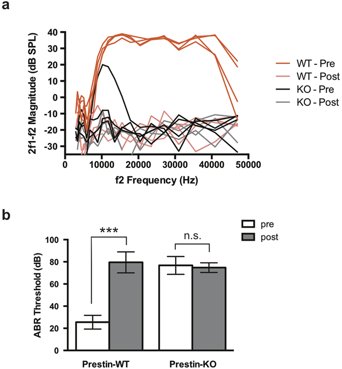
Figure 2. In vivo physiology of HPβCD-treated WT- and prestin-KO mice.

(a) Distortion product otoacoustic emissions (DPOAEs) for WT and prestin-KO mice (n = 4 each) on the FVB background were recorded for L1 = L2 = 70 dB SPL before (“Pre”) and 7 days after (“Post”) 8000 mg/kg HPβCD treatment as in Fig. 1. HPβCD treatment abolishes DPOAEs in WT mice (faint red traces). Because prestin-KO mice (black traces) already have reduced emissions (Liberman et al., 2002), HPβCD did not affect DPOAEs in these mice as dramatically as in controls. (b) Auditory brainstem responses (ABR) from the same WT and prestin-KO mice as in (a) were determined using a 32 kHz tone burst before (“Pre”) and after (“Post”) HPβCD treatment. HPβCD treatment significantly increased the ABR threshold in WT mice. Prestin-KO mice already have a large threshold shift (Liberman et al., 2002) so that HPβCD did not affect ABR thresholds in these mice. Mean ± SD are plotted; significance was determined using an unpaired t-test. n.s., not significant; ***p ≤ 0.0001.
