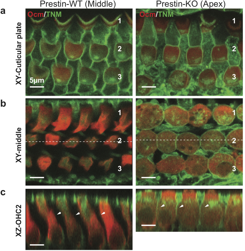
Figure 3. Cholesterol staining of cochleae from WT- and prestin-KO mice.
Saline-treated WT- and prestin-KO cochleae (as in Fig. 1) were stained with TNM-fluorescein (TNM-FL) and imaged. (a) Single plane image at the cuticular plate (XY view, “Cuticular plate”) and (b) along the lateral wall (XY view, “middle”). Both WT and KO cochleae show similar cholesterol staining (green) for all rows of OHCs at the cuticular plate. (c) Radial view of the z-stack images. Unlike the KO, staining was not observed on the lateral walls of OHCs from WT cochlea (XZ view, “OHC-2”, white arrowheads). Red, anti-oncomodulin; Green, TNM-FL. Scale bars, 5 μm.

