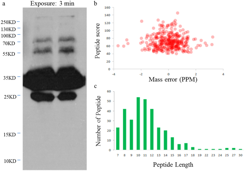
Figure 1. QC validation of LC-MS/MS data.
(a) Western blotting analysis of 5102WB with pan anti-succinylation antibody demonstrates the presence of succinylated proteins. In total, 40 μg of whole tissue lysate was loaded in one lane, and primary antibody was diluted by 1:1000. (b) The distribution of mass error. (c) T-distribution of succinylated peptides based on their length.

