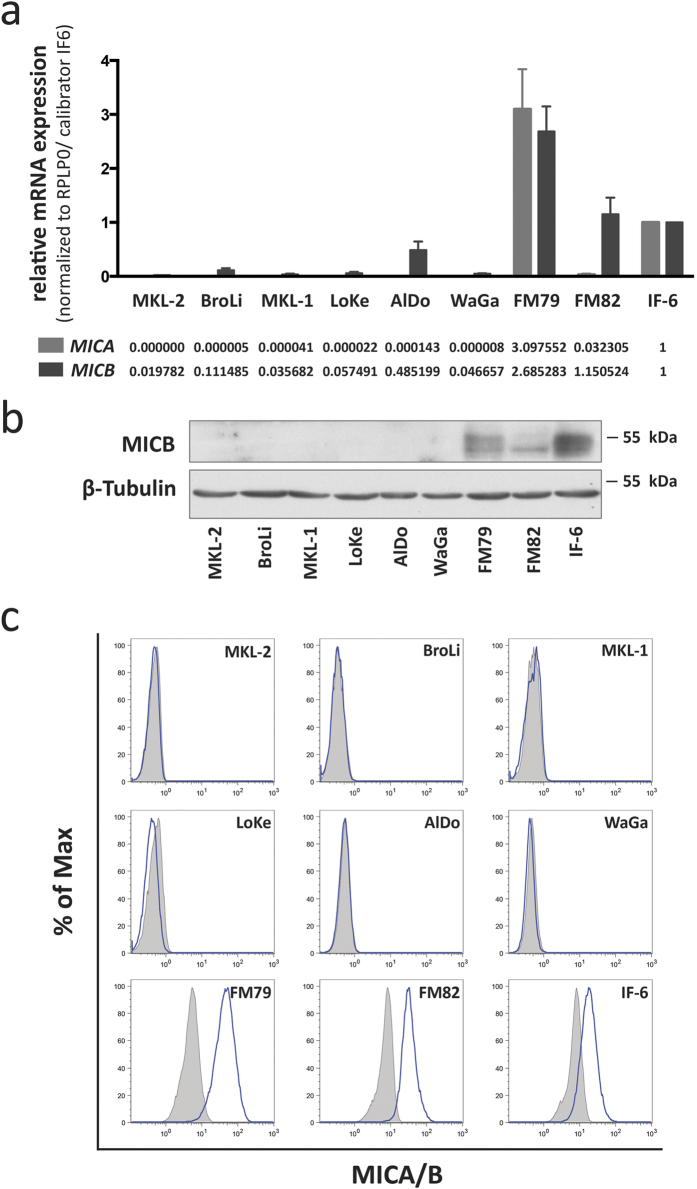
Figure 2. MCC cell lines do not express MICA and MICB protein despite low levels of MICA and MICB mRNA.
(a) MICA (light grey) and MICB (dark grey) mRNA expression was determined by qRT-PCR in MCC cells. Relative expression levels were calculated by normalization of CT values to RPLP0 and calibration to the melanoma cell line IF6. (b) MICB protein expression of whole cell lysates was determined by immunoblot; β-tubulin served as a loading control. (c) MICA/B cell surface expression was determined by flow cytometry using an antibody recognizing both MICA and MICB (clone 6D4; blue line); matched isotype control is depicted as grey filled area. Melanoma cell lines FM79, FM82 and IF6, served as positive control for MICA and MICB expression in all assays illustrated in this figure.

