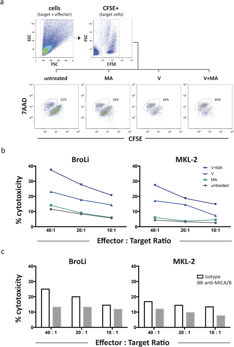
Figure 5. Inhibition of HDACs in MCC cell lines increased their susceptibility to LAK cell mediated lysis, which is is subdued by MICA/B blockade.
The flow cytometry based cytotoxicity assay was performed as described in Material and Methods. (a) The gating strategy is illustrated for untreated and treated BroLi cells used at an effector to target ratio of 40:1; target cells were gated as CFSE positive cells in an FSC/CFSE plot, lysed target cells were defined as 7AAD/CFSE double positive cells and are quantified as percentage of all target cells. (b) Untreated (grey), vorinostat (V, light blue), mithramycin A (MA, turquoise), or the combination thereof (V+MA, dark blue) treated BroLi and MKL-2 cells served as target cells for LAK cells at the indicated effector to target ratios in a 4h cytotoxicity assay. The lysis of the respective target is given as average of three independent experiments. (c) Vorinostat plus mithramycin A treated BroLi and MKL-2 cells served as target cells for LAK cells at the indicated effector to target ratios in a 4h cytotoxicity assay in the presence of saturating amounts of a MICA/B specific blocking antibody (grey bars) or an isotype control antibody (white bars); Fc receptors of effector cells were blocked by saturating amounts of F(ab)2 fragments.

