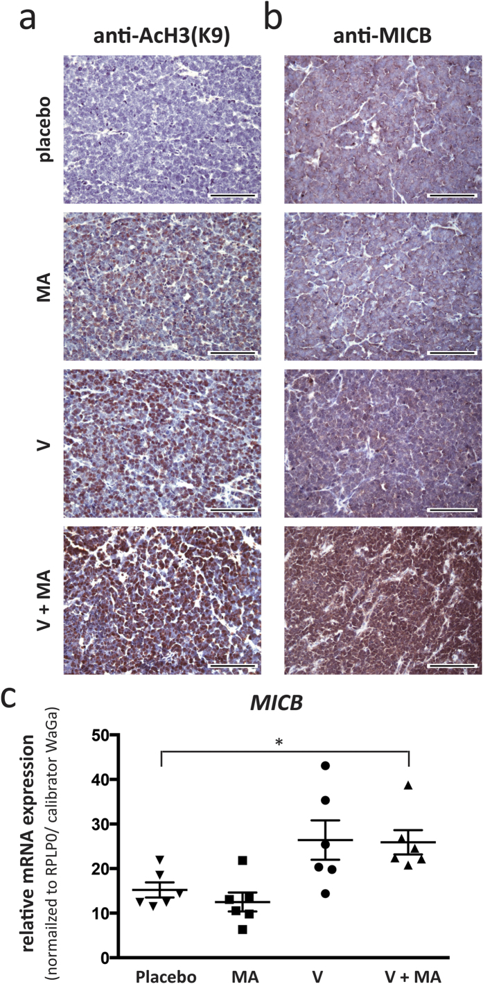
Figure 6. Vorinostat and mithramycin A treatment induces histone H3K9 acetylation and MICB expression in MCC cells in vivo.
NOD.CB17/Prkdcscid mice (n = 6 for each treatment group) bearing subcutaneous xenotransplants of WaGa cells were treated with placebo, vorinostat (V) mithramycin A (MA), or the combination thereof (V+MA) as described in materials and methods. Immunohistochemistry on FFPE fixed tumor samples obtained after two weeks of treatment was performed using antibodies specific against AcH3K9 (a) or MICB (b). Representative examples are depicted at 40× magnification, scale bar is 100 μm. (c) mRNA was isolated from cryopreserved tumors and qRT-PCR was performed using primers specific for MICB. CT values were normalized to RPLP0 and calibrated to in vitro cultured WaGa cells. Statistical analysis was performed using the Kruskal-Wallis test.

