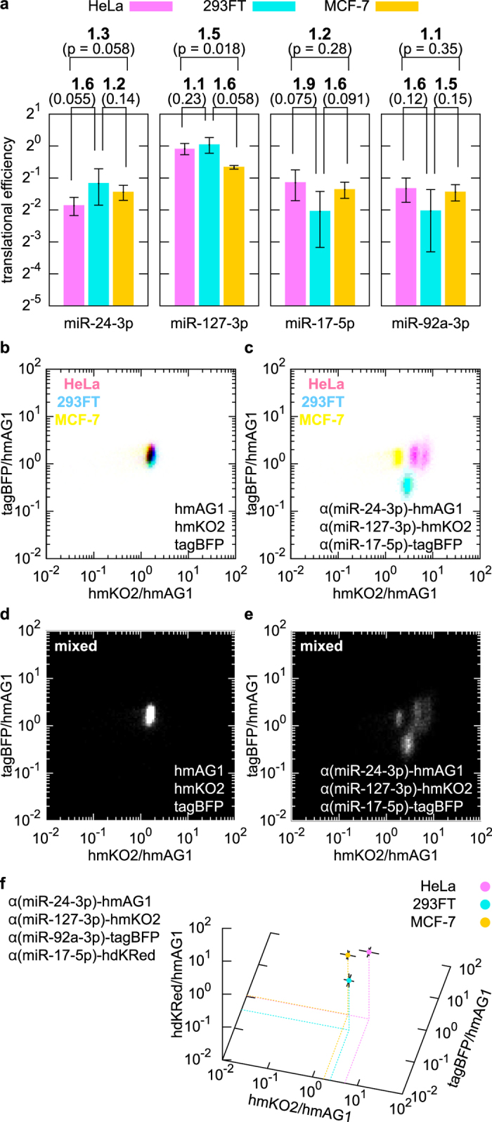
Figure 3. High-resolution identification and separation of three cell types using three miRNA-responsive mRNAs.

(a) Translational efficiency of three miRNA-responsive mRNAs in HeLa (magenta), 293FT (cyan), and MCF-7 (yellow) cells, shown as in Fig. 2b. (b,c) 2-D densities of HeLa, 293FT and MCF-7 cells individually transfected with either control reporter mRNAs (b) or the three indicated miRNA-responsive mRNAs (c). The flow cytometry data are plotted against two fluorescence ratios: hmKO2 or tagBFP intensity divided by hmAG1 intensity. (d,e) Separation of mixed cells in 2-D space. The three lines were cocultured and transfected with the same mRNA set as in (b) or (c). (f) 3-D separation using four miRNA-responsive mRNAs. The geometric mean values of three ratios (intensities of hmKO2, tagBFP or hdKRed divided by the intensity of hmAG1 in each cell) were calculated and plotted. The distribution of the cells is shown in Supplementary Fig. S7.
