. 2015 Oct-Dec;38(4):420–432. doi: 10.1590/S1415-475738420150019

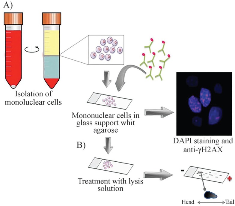
Figure 2. General assays for detecting DNA damage (A) Immunohistochemistry with antibodies directed against γ-H2AX: peripheral blood mononuclear cells are isolated, nuclei are stained with DAPI and with antibodies directed at γ-stained H2AX and visualized under fluorescent microscopy. (B) Comet assay: the comet assay is also performed on mononuclear cells. The cells are embedded in agarose on a thin glass slide, cells are lysed and incubated in an alkaline solution. Subsequently, DNA fragments are separated by electrophoresis and stained with ethidium bromide. The comet-like image is viewed under a fluorescence microscope. The length of the comet tail indicates the frequency of DNA breaks.

Figure 2